 Use cases
About 130 years after its discovery, Tuberculosis remains a persistent threat and a leading cause of death worldwide according to WHO. Generally, TB can be cured with antibiotics. However, the different types of TB require different treatments, and therefore the detection of the TB type and the evaluation of lesion characteristics are important real-world tasks.
ImageClef task - automated CT report generation: presence of TB lesions in general, presence of pleurisy and caverns in particular.
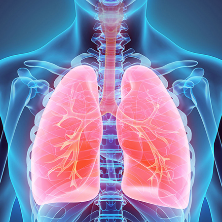
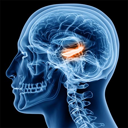
Gliomas are the most common primary brain malignancies, with different degrees of aggressiveness, variable prognosis and various heterogeneous histological sub-regions, i.e., peritumoral edema, necrotic core, enhancing and non-enhancing tumor core. Due to this highly heterogeneous appearance and shape, segmentation of brain tumors in multimodal MRI scans is one of the most challenging tasks in medical image analysis.
BRATS 2020 tasks: produce segmentation labels of the different glioma sub-regions (the "enhancing tumor", the "tumor core" and the "whole tumor") and predict patient overall survival.
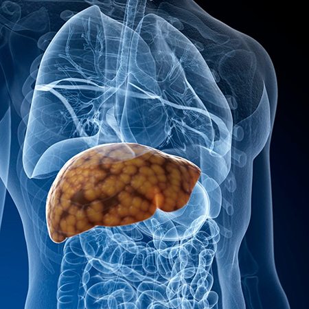
Development of a software capable of detecting the presence of hepatocarcinoma in CTs.
The typical protocol at IRO includes 4 acquisitions (scans):
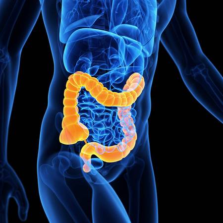
Colorectal cancer is the third most commonly diagnosed cancer in men and women. Since tumor stage at time of diagnosis is a critical determinant of patient outcome, early detection of colorectal cancer by screening modalities holds the key to improving patient survival.
IRO tasks: identify colorectal tumors in MRIs; analyze the evolution during radio-therapy.
The reduced blood flow (stalled vessels) in the brain is associated with Alzheimer’s disease and other forms of dementia, but until recently no one knew why. Researches from Cornell University have developed new imaging techniques which might tell them the reason, but they need to speed up the detection of stalled vessels. Using artificial intelligence, may speed up their research by 10 times.
Clog Loss tasks: Using half of million of annotated two-photon excitation microscopy videos, find the stalled vessels for a very unbalanced dataset.
